General Medical Semiology Guide Part II 1st Edition by Manuela Stoicescu 0128220382 9780128220382
$50.00 Original price was: $50.00.$25.00Current price is: $25.00.
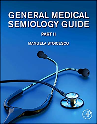
General Medical Semiology Guide Part II 1st Edition by Manuela Stoicescu – Ebook PDF Instant Download/DeliveryISBN: 0128220382, 9780128220382
Full download General Medical Semiology Guide Part II 1st Edition after payment.
Product details:
ISBN-10 : 0128220382
ISBN-13 : 9780128220382
Author : Manuela Stoicescu
General Medical Semiology Guide, Part Two is the second volume in a two volume set that provides a comprehensive understanding of medical semiology. Highly illustrated with many original images from the author’s daily medical practice, the book highlights all signs of diseases and important semiological maneuvers. Each chapter contains a specific questionnaire of important questions that should be asked of patients in different situations to obtain valuable information that will assist in both medical thinking and in the formulation of diagnoses. Part Two covers topics on how to examine primary and secondary skin lesions, hair changes, nails, lymph nodes, breasts, and more.
General Medical Semiology Guide Part II 1st Table of contents:
Chapter 1. Skin Lesions
1.1. Primary Skin Lesions
1.2. Secondary Skin Lesions
Chapter 2. Changes in Hair
2.1. Hypotrichosis
2.2. Hypertrichosis
Hypertrichosis
Chapter 3. The Nails
3.1. Clubbed Fingers
3.1. Clubbed Fingers
3.1. Clubbed Fingers
3.2. Hypertrophic Osteoarthropathy, Bamberger–Marie Disease
3.3. Spoon Nails—Koilonychia
3.3. Spoon Nails—Koilonychia
3.4. Mycosis of the Nails—Onychomycosis
3.5. The Appearance of the Nails After Frostbite
3.6. Nail Changes in Peripheral Arterial Disease
3.7. Onychomycosis in Patients With Cardiac Failure
Chapter 4. Nutritional Status
4.1. Obesity
4.2. Cushing Syndrome
4.3. Abdominal Obesity and White Stretch Marks
4.4. Cachexia
Chapter 5. Subcutaneous Edema
5.1. Indentations—Pitting Edema
5.2. Increased Hydrostatic Pressure in the Capillary
5.3. Decreased Colloid Osmotic (Oncotic) Pressure in the Capillary
5.4. Colloid Osmotic (Oncotic) Pressure increases in the Interstitial Space Because of Obstruction by the protein-rich Lymphatic System
5.5. Generalized Edema
5.6. Ascites and Orange Peel Sign
5.6. Ascites and Orange Peel Sign
5.7. Orange Peel Sign Suggests Edema of the Abdominal Wall
5.8. Ascites—Orange Peel Sign
5.8. Ascites—Orange Peel Sign
5.8. Ascites—Orange Peel Sign
5.8. Ascites—Orange Peel Sign
5.8. Ascites—Orange Peel Sign
5.9. Edema of the Lower Limbs
5.10. Edema of the Lower Limbs in the Same Patient—Indentation
5.11. Cardiac Edema
5.11. Cardiac Edema
5.12. Clinical Case Presentation
5.13. Stasis Dermatitis Pigmentation
5.14. Renal Edema
5.15. Hepatic Edema
5.15. Hepatic Edema
5.16. Endocrine Edema
5.17. Localized Edema
Chapter 6. Collateral Circulation
6.1. Cyanosis—Edema in Cape
6.2. Arterial Collateral Circulation
6.3. Venous Collateral Circulation
6.3. Venous Collateral Circulation
Chapter 7. Lymph Node System
7.1. Superficial Lymph Node System
7.2. Deep Lymph Node System
7.3. Adenomegaly
7.4. Mediastinal Lymph Nodes
7.4. Mediastinal Lymph Nodes
7.5. Abdominal Lymph Node
7.6. The Objective Examination
Chapter 8. The Osteoarticular System
8.1. Hand Like the Back of a Camel—Rheumatoid Arthritis
8.1. Hand like the Back of a Camel—Rheumatoid Arthritis
8.2. Rheumatoid Nodules in a Young Girl Patient With Rheumatoid Arthritis
8.3. Dupuytren Disease
8.3. Dupuytren Disease
8.3. Dupuytren Disease
8.3. Dupuytren Disease
8.3. Dupuytren Disease
8.3. Dupuytren Disease
8.4. Nodules of Arthrosis—Heberden and Bouchard
8.4. Nodules of Arthrosis—Heberden and Bouchard
8.4. Nodules of Arthrosis—Heberden and Bouchard
8.4. Nodules of Arthrosis—Heberden and Bouchard
8.5. Comparison Between Nodules of Arthrosis—Heberden and Bouchard
8.6. Nodules of Arthrosis—Heberden and Bouchard
8.7. Bouchard Nodules of Arthrosis
8.6. Nodules of Arthrosis—Heberden and Bouchard
8.6. Nodules of Arthrosis—Heberden and Bouchard
8.7. Bouchard Nodules of Arthrosis
8.8. The fifth Finger Fixed in Flexion—Sequela of Arthrosis
8.6. Nodules of Arthrosis—Bouchard and Heberden
8.7. Bouchard Nodules of Arthrosis
8.9. Gouty Tophi
8.9. Gouty Tophi
8.9. Gouty Tophi
8.9. Gouty Tophi
8.9. Gouty Tophi
8.9. Gouty Tophi
8.9. Gouty Tophi
8.9. Gouty Tophi
8.9. Gouty Tophi
8.10. Gouty Tophi—Knees
8.10. Gouty Tophi—Knees
8.10. Gouty Tophi—Knees
8.11. Gouty Tophi Interphalangeal Joints and Metacarpophalangeal Joints
8.12. Gouty Tophi—Elbow
8.12. Gouty tophus—Elbow
8.12. Gouty tophus—Elbow
8.12. Gouty tophus—Elbow
8.12. Gouty Tophi—Elbow
8.13. Gouty Tophi—Ear Pinna
8.14. Gouty Tophi—Interphalangeal Joints
8.15. Another Alcoholic Patient with Chronic Gouty Tophi—Ear Pinna
8.16. Gouty Tophi—Ear Pinna
8.16. Gouty Tophi—Ear Pinna
8.17. Acute Attack of Gout
8.17. Acute Attack of Gout
8.18. Gouty Tophi—Amputation of some Fingers
8.19. Swelling of the Interphalangeal Joint
8.20. Gouty Tophi—Amputation of some Fingers
8.21. Gouty Tophi
8.21. Gouty Tophi
8.22. Clinical Case No. 1
8.23. Clinical Case no. 2–Acute Gout Attack
8.24. The Objective Examination of the Knees
8.25. Ligamentous Hyperlaxity of the Interphalangeal Joint—Marfan Syndrome
Chapter 9. Fever
9.1. Body Temperature Measurement
Chapter 10. The Semiology of the Breast
10.1. Questionnaire
10.2. The Topographic division of the Breast into Four Quadrants
10.3. The Objective Examination of the Breast
People also search for General Medical Semiology Guide Part II 1st:
borrow general medical semiology guide part i
general medical semiology guide part i pdf
general medical semiology guide pdf
general medical check-up list
what does a general medical exam include
Tags: General Medical, Semiology Guide, Manuela Stoicescu, specific questionnaire
You may also like…
Politics & Philosophy - Social Sciences
Science (General)
Medicine
Politics & Philosophy
Power in Economic Thought 1st Edition by Manuela Mosca 3030067807 978-3030067809
Reference - School Guides & Test Preparation
CUET-UG : General Test (Section-III) Exam Guide 1st Edition Rph Editorial Board


