(Ebook PDF) Breast Imaging The Requisites 3rd Edition by Debr Ikeda, Kanae Kawai Miyake 0323391567 9780323391566 full chapters
$50.00 Original price was: $50.00.$25.00Current price is: $25.00.
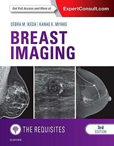
Breast Imaging: The Requisites 3rd Edition by Debra M. Ikeda, Kanae Kawai Miyake – Ebook PDF Instant Download/DeliveryISBN: 0323391567, 9780323391566
Full download Breast Imaging: The Requisites 3rd Edition after payment.
Product details:
ISBN-10 : 0323391567
ISBN-13 : 9780323391566
Author : Debra M. Ikeda, Kanae Kawai Miyake
Now in its 3rd Edition, this bestselling volume in the popular Requisites series, by Drs. Debra M. Ikeda and Kanae K. Miyake, thoroughly covers the fast-changing field of breast imaging. Ideal for residency, clinical practice and certification and MOC exam study, it presents everything you need to know about diagnostic imaging of the breast, including new BI-RADS standards, new digital breast tomosynthesis (DBT) content and videos, ultrasound, and much more. Compact and authoritative, it provides up-to-date, expert guidance in reading and interpreting mammographic, ultrasound, DBT, and MRI images for efficient and accurate detection of breast disease.
Breast Imaging: The Requisites 3rd Table of contents:
Chapter 1. Mammography Acquisition: Screen-Film, Digital Mammography and Tomosynthesis, the Mammography Quality Standards Act, and Computer-Aided Detection
Technical Aspects of Mammography Image Acquisition
Image Evaluation and Artifacts
Quality Assurance in Mammography and the Mammography Quality Standards Act
Computer-Aided Detection
Conclusion
Chapter 2. Mammogram Analysis and Interpretation
Breast Cancer Risk Factors
Signs and Symptoms of Breast Cancer
The Normal Mammogram
Mammographic Findings of Breast Cancer
Display of Mammograms
Location of a Finding
Systematic Approach to Mammography Interpretation
Reporting of Mammographic Findings Based on BI-RADS 2013
Diagnostic Mammography
Diagnostic Limitations
Chapter 3. Mammographic Analysis of Breast Calcifications
Normal Breast Anatomy and Calcifications
Breast Ducts, Terminal Ductal Lobular Units, Pathology, and Calcifications
Technique for Finding Calcifications
Calcification Classifications Based on Breast Imaging Reporting and Data System 2013
Stable Suspicious Calcifications
Breast Imaging Reporting and Data System Category 3 Calcifications (Grouped Punctate) and Management
Ultrasound in Evaluation of Calcifications
Ductal Carcinoma in Situ Appearance on Mammography Versus Magnetic Resonance Imaging
Chapter 4. Mammographic and Ultrasound Analysis of Breast Masses
Mammography for Evaluating Nonpalpable Masses, Asymmetries, and Palpable Masses
Description of Masses, Asymmetry, and Architectural Distortion Based on Breast Imaging Reporting and Data System 2013 Mammography
Further Characterization of Masses with Ultrasonography
Mammographic and Ultrasound Atlas of Masses
Chapter 5. Breast Ultrasound Principles
Technical Considerations
Breast Anatomy
Ultrasound Lexicon in Breast Imaging Reporting and Data System for Ultrasound (2013)
Masses: Feature Analytic Approach
Appropriateness Criteria, American College of Radiology Practice Parameters, and Other Guidelines
Special Indications
Chapter 6. Mammographic and Ultrasound-Guided Breast Biopsy Procedures
Before Procedure
Percutaneous Needle Biopsy of Cysts, Solid Masses, or Calcifications
Preoperative Needle Localization
Chapter 7. Magnetic Resonance Imaging of Breast Cancer and Magnetic Resonance Imaging–Guided Breast Biopsy
Magnetic Resonance Imaging Techniques
Interpretation of Breast Magnetic Resonance Imaging
Breast Magnetic Resonance Imaging Atlas
Indications
Magnetic Resonance Imaging–Directed Intervention
Chapter 8. Breast Cancer Treatment-Related Imaging and the Postoperative Breast
Combined Clinical and Imaging Workup of Breast Abnormalities
Breast Cancer Diagnosis and Treatment
Evaluation of Axillary Lymph Nodes
Clinical and Breast Imaging Factors in Determining Appropriate Local Therapy: Lumpectomy or Mastectomy
Preoperative Imaging
Normal Postoperative Imaging Changes After Breast Biopsy or Lumpectomy
Whole-Breast, External Beam Radiotherapy, and Accelerated Partial Breast Irradiation
Breast Imaging Before Reexcision Lumpectomy or Radiotherapy
Normal Imaging Changes After Radiation Therapy
Treatment Failure or Ipsilateral Breast Tumor Recurrence (IBTR)
Mastectomy
Breast Imaging for Assessing Response to Neoadjuvant Chemotherapy
Chapter 9. Breast Implants and the Reconstructed Breast
Breast Implants
Mammography and Implants
Implant Complications
Imaging Evaluation of Implant Rupture
Direct Silicone/Paraffin Injection
Breast Reconstruction
Reduction Mammoplasty
Chapter 10. Clinical Breast Problems and Unusual Breast Conditions
The Male Breast: Gynecomastia and Male Breast Cancer
Pregnant and Lactating Patients and Pregnancy-Associated Breast Cancer
Probably Benign Findings (Breast Imaging Reporting and DATA System Category 3)
Nipple Discharge and Galactography
Nipple and Skin Retraction
Breast Edema
Hormone Changes
Breast Pain
Axillary Lymphadenopathy
Paget Disease of the Nipple
Sarcomas
Mondor Disease
Granulomatous Mastitis
Diabetic Mastopathy
Desmoid Tumor
Trichinosis
Dermatomyositis
Foreign Bodies
Hidradenitis Suppurativa
Neurofibromatosis
Lymphangioma
Poland Syndrome
Chapter 11. 18F-FDG PET/CT and Nuclear Medicine for the Evaluation of Breast Cancer
PET/CT Scanning Principles
Clinical Utility of 18F-Fluorodeoxyglucose Position Emission Tomography and Position Emission Tomography/Computed Tomography for Breast Cancer
Developing Positron Emission Tomography Technologies
Conventional Nuclear Medicine Techniques for the Diagnosis of Breast Cancer
Conclusions
People also search for Breast Imaging: The Requisites 3rd:
breast imaging the core requisites 4th edition
what is a breast imaging
what is a breast imaging exam
what does breast imaging mean
breast imaging procedures
Tags:
Breast Imaging,The Requisites,Debr Ikeda,Kanae Kawai Miyake
You may also like…
Medicine - Pediatrics
Diagnostic Imaging: Pediatric Neuroradiology 3rd Edition Kevin R. Moore
Medicine - Radiology
Emergency radiology the requisites 2nd Edition by Jorge Soto, Brian Lucey 0323340865 9780323340861
Medicine - Cardiology
Medicine - Oncology
Medicine - Radiology
Nuclear Medicine and Molecular Imaging: the Requisites 5th Edition Janis P. O’Malley
Uncategorized
Medicine - Others


