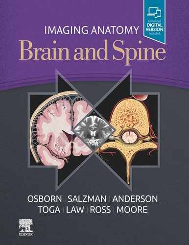
(Ebook PDF) Imaging Anatomy Brain and Spine 1st edition by Anne Osborn 0323661157 9780323661157 full chapters
$50.00 Original price was: $50.00.$25.00Current price is: $25.00.
Imaging Anatomy Brain and Spine 1st edition by Anne G. Osborn – Ebook PDF Instant Download/DeliveryISBN: 0323661157, 9780323661157
Full download Imaging Anatomy Brain and Spine 1st edition after payment.
Product details:
ISBN-10 : 0323661157
ISBN-13 : 9780323661157
Author : Anne G. Osborn
This richly illustrated and superbly organized text/atlas is an excellent point-of-care resource for practitioners at all levels of experience and training. Written by global leaders in the field, Imaging Anatomy: Brain and Spine provides a thorough understanding of the detailed normal anatomy that underlies contemporary imaging. This must-have reference employs a templated, highly formatted design; concise, bulleted text; and state-of- the-art images throughout that identify the clinical entities in each anatomic area.
Imaging Anatomy Brain and Spine 1st Table of contents:
Part I: Brain
SECTION 1: SCALP, SKULL, AND MENINGES
Chapter 1: Scalp and Calvarial Vault
Main Text
Image Gallery
Chapter 2: Cranial Meninges
Main Text
Image Gallery
Chapter 3: Pia and Perivascular Spaces
Main Text
Image Gallery
SECTION 2: SUPRATENTORIAL BRAIN ANATOMY
Chapter 4: Cerebral Hemispheres Overview
Main Text
Image Gallery
Chapter 5: Gyral/Sulcal Anatomy
Main Text
Image Gallery
Chapter 6: White Matter Tracts
Main Text
Image Gallery
Chapter 7: Basal Ganglia and Thalamus
Main Text
Image Gallery
Chapter 8: Other Deep Gray Nuclei
Main Text
Image Gallery
Chapter 9: Limbic System
Main Text
Image Gallery
Chapter 10: Sella, Pituitary, and Cavernous Sinus
Main Text
Image Gallery
Chapter 11: Pineal Region
Main Text
Image Gallery
Chapter 12: Primary Somatosensory Cortex (Areas 1, 2, 3)
Main Text
Image Gallery
Chapter 13: Primary Motor Cortex (Area 4)
Main Text
Image Gallery
Chapter 14: Superior Parietal Cortex (Areas 5, 7)
Main Text
Image Gallery
Chapter 15: Premotor Cortex and Supplementary Motor Area (Area 6)
Main Text
Image Gallery
Chapter 16: Superior Prefrontal Cortex (Area 8)
Main Text
Image Gallery
Chapter 17: Dorsolateral Prefrontal Cortex (Areas 9, 46)
Main Text
Image Gallery
Chapter 18: Frontal Pole (Area 10)
Main Text
Image Gallery
Chapter 19: Orbitofrontal Cortex (Area 11)
Main Text
Image Gallery
Chapter 20: Insula and Parainsula Areas (Areas 13, 43)
Main Text
Image Gallery
Chapter 21: Primary Visual and Visual Association Cortex (Areas 17, 18, 19)
Main Text
Image Gallery
Chapter 22: Temporal Cortex (Areas 20, 21, 22)
Main Text
Image Gallery
Chapter 23: Posterior Cingulate Cortex (Areas 23, 31)
Main Text
Image Gallery
Chapter 24: Anterior Cingulate Cortex (Areas 24, 32, 33)
Main Text
Image Gallery
Chapter 25: Subgenual Cingulate Cortex (Area 25)
Main Text
Image Gallery
Chapter 26: Retrosplenial Cingulate Cortex (Areas 29, 30)
Main Text
Image Gallery
Chapter 27: Parahippocampal Gyrus (Areas 28, 34, 35, 36)
Main Text
Image Gallery
Chapter 28: Fusiform Gyrus (Area 37)
Main Text
Image Gallery
Chapter 29: Temporal Pole (Area 38)
Main Text
Image Gallery
Chapter 30: Inferior Parietal Lobule (Areas 39, 40)
Main Text
Image Gallery
Chapter 31: Primary Auditory and Auditory Association Cortex (Areas 41, 42)
Main Text
Image Gallery
Chapter 32: Inferior Frontal Gyrus (Areas 44, 45, 47)
Main Text
Image Gallery
SECTION 3: BRAIN NETWORK ANATOMY
Chapter 33: Functional Network Overview
Main Text
Image Gallery
Chapter 34: Neurotransmitter Systems
Main Text
Image Gallery
Chapter 35: Default Mode Network
Main Text
Image Gallery
Chapter 36: Attention Control Network
Main Text
Image Gallery
Chapter 37: Sensorimotor Network
Main Text
Image Gallery
Chapter 38: Visual Network
Main Text
Image Gallery
Chapter 39: Limbic Network
Main Text
Image Gallery
Chapter 40: Language Network
Main Text
Image Gallery
Chapter 41: Memory Network
Main Text
Image Gallery
Chapter 42: Social Network
Main Text
Image Gallery
SECTION 4: INFRATENTORIAL BRAIN
Chapter 43: Brainstem and Cerebellum Overview
Main Text
Image Gallery
Chapter 44: Midbrain
Main Text
Image Gallery
Chapter 45: Pons
Main Text
Image Gallery
Chapter 46: Medulla
Main Text
Image Gallery
Chapter 47: Cerebellum
Main Text
Image Gallery
Chapter 48: Cerebellopontine Angle/IAC
Main Text
Image Gallery
SECTION 5: CSF SPACES
Chapter 49: Ventricles and Choroid Plexus
Main Text
Image Gallery
Chapter 50: Subarachnoid Spaces/Cisterns
Main Text
Image Gallery
SECTION 6: SKULL BASE AND CRANIAL NERVES
Chapter 51: Skull Base Overview
Main Text
Image Gallery
Chapter 52: Anterior Skull Base
Main Text
Image Gallery
Chapter 53: Central Skull Base
Main Text
Image Gallery
Chapter 54: Posterior Skull Base
Main Text
Image Gallery
Chapter 55: Cranial Nerves Overview
Main Text
Image Gallery
Chapter 56: Olfactory Nerve (CNI)
Main Text
Image Gallery
Chapter 57: Optic Nerve (CNII)
Main Text
Image Gallery
Chapter 58: Oculomotor Nerve (CNIII)
Main Text
Image Gallery
Chapter 59: Trochlear Nerve (CNIV)
Main Text
Image Gallery
Chapter 60: Trigeminal Nerve (CNV)
Main Text
Image Gallery
Chapter 61: Abducens Nerve (CNVI)
Main Text
Image Gallery
Chapter 62: Facial Nerve (CNVII)
Main Text
Image Gallery
Chapter 63: Vestibulocochlear Nerve (CNVIII)
Main Text
Image Gallery
Chapter 64: Glossopharyngeal Nerve (CNIX)
Main Text
Image Gallery
Chapter 65: Vagus Nerve (CNX)
Main Text
Image Gallery
Chapter 66: Accessory Nerve (CNXI)
Main Text
Image Gallery
Chapter 67: Hypoglossal Nerve (CNXII)
Main Text
Image Gallery
SECTION 7: EXTRACRANIAL ARTERIES
Chapter 68: Aortic Arch and Great Vessels
Main Text
Image Gallery
Chapter 69: Cervical Carotid Arteries
Main Text
Image Gallery
SECTION 8: INTRACRANIAL ARTERIES
Chapter 70: Intracranial Arteries Overview
Main Text
Image Gallery
Chapter 71: Intracranial Internal Carotid Artery
Main Text
Image Gallery
Chapter 72: Circle of Willis
Main Text
Image Gallery
Chapter 73: Anterior Cerebral Artery
Main Text
Image Gallery
Chapter 74: Middle Cerebral Artery
Main Text
Image Gallery
Chapter 75: Posterior Cerebral Artery
Main Text
Image Gallery
Chapter 76: Vertebrobasilar System
Main Text
Image Gallery
SECTION 9: VEINS AND VENOUS SINUSES
Chapter 77: Intracranial Venous System Overview
Main Text
Image Gallery
Chapter 78: Dural Sinuses
Main Text
Image Gallery
Chapter 79: Superficial Cerebral Veins
Main Text
Image Gallery
Chapter 80: Deep Cerebral Veins
Main Text
Image Gallery
Chapter 81: Posterior Fossa Veins
Main Text
Image Gallery
Chapter 82: Extracranial Veins
Main Text
Image Gallery
Part II: Spine
SECTION 1: VERTEBRAL COLUMN, DISCS, AND PARASPINAL MUSCLE
Chapter 83: Vertebral Column Overview
Main Text
Image Gallery
Chapter 84: Ossification
Main Text
Image Gallery
Chapter 85: Vertebral Body and Ligaments
Main Text
Image Gallery
Chapter 86: Intervertebral Disc and Facet Joints
Main Text
Image Gallery
Chapter 87: Paraspinal Muscles
Main Text
Image Gallery
Chapter 88: Craniocervical Junction
Main Text
Image Gallery
Chapter 89: Cervical Spine
Main Text
Image Gallery
Chapter 90: Thoracic Spine
Main Text
Image Gallery
Chapter 91: Lumbar Spine
Main Text
Image Gallery
Chapter 92: Sacrum and Coccyx
Main Text
Image Gallery
SECTION 2: CORD, MENINGES, AND SPACES
Chapter 93: Spinal Cord and Cauda Equina
Main Text
Image Gallery
Chapter 94: Meninges and Compartments
Main Text
Image Gallery
SECTION 3: VASCULAR
Chapter 95: Spinal Arterial Supply
Main Text
Image Gallery
Chapter 96: Spinal Veins and Venous Plexus
Main Text
Image Gallery
SECTION 4: PLEXI AND PERIPHERAL NERVES
Chapter 97: Brachial Plexus
Main Text
Image Gallery
Chapter 98: Lumbar Plexus
Main Text
Image Gallery
Chapter 99: Sacral Plexus and Sciatic Nerve
Main Text
Image Gallery
Chapter 100: Peripheral Nerve and Plexus Overview
People also search for Imaging Anatomy Brain and Spine 1st:
does a spine mri show organs
what does a brain and spine mri show
is the spine connected to the skull
what does an mri of the spine look like
brain imaging anatomy
Tags:
Imaging,Anatomy Brain,Spine,Anne Osborn
You may also like…
Medicine Medicine - Others
Medicine - Others
Uncategorized
Medicine - Neurology
Essentials of Osborn’s Brain: A Fundamental Guide for Residents and Fellows 1st Edition


