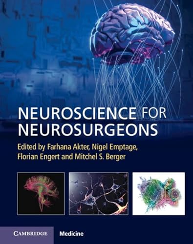
Neuroscience for Neurosurgeons 1st Edition by Farhana Akter 1108918468 9781108918466
$50.00 Original price was: $50.00.$25.00Current price is: $25.00.
Neuroscience for Neurosurgeons 1st Edition by Farhana Akter – Ebook PDF Instant Download/DeliveryISBN: 1108918468, 9781108918466
Full download Neuroscience for Neurosurgeons 1st Edition after payment.
Product details:
ISBN-10 : 1108918468
ISBN-13 : 9781108918466
Author : Farhana Akter
With advances in medicine and medical innovation, the face of neurosurgery has changed dramatically. A new era of surgeons value the need to undertake research in everyday practice and actively participate in the clinic and laboratory in order to improve patient prognosis. Highlighting the principles of basic neuroscience and its application to neurosurgical disease, this book breaks down neurological conditions into current academic themes and advances. The book is split into two sections, with the first covering basic and computational neuroscience including neuroanatomy, neurophysiology, and the growing use of artificial intelligence. The second section concentrates on specific conditions, such as gliomas, degenerative cervical myelopathy and peripheral nerve injury. Outlining the pathophysiological underpinnings of neurosurgical conditions and the key investigative tools used to study disease burden, this book will be an invaluable source for the academic neurosurgeon undertaking basic and translational research.
Neuroscience for Neurosurgeons 1st Table of contents
Section 1 Basic and Computational Neuroscience
Chapter 1 Neuroanatomy
1.1 Anatomical Planes and Orientation of the Brain
1.2 Vascular Supply to the Brain
1.2.1 Circle of Willis
1.2.2 Cerebral Venous Drainage
1.2.3 Lymphatics of the Brain
1.3 Bones of the Skull
1.4 The Cranial Fossae
1.5 Layers of the Scalp
1.6 Meninges
1.6.1 Dura Mater
1.6.2 Arachnoid Mater
1.6.3 Pia Mater
1.7 Organization of the Sympathetic and Parasympathetic Nervous Systems
1.7.1 Sympathetic Ganglia Supplying the Head and Neck: Cervical Ganglia
1.7.2 Parasympathetic Ganglia Supplying the Head and Neck
1.8 Structures of the Brain
1.9 Forebrain
1.10 Cerebrum
1.10.1 Brodmann’s Map
1.10.2 Layers of The Cerebral Cortex
1.10.3 Vascular Supply
1.11 Ventricles
1.12 Higher Association Areas of the Cortex
1.13 Limbic System
1.13.1 Basal Ganglia
1.13.1.1 Connections of the Basal Ganglia
1.13.2 Thalamus
1.13.3 Cingulate Gyrus
1.13.4 Hippocampus
1.13.4.1 Dentate Gyrus
1.13.4.2 Hippocampal Inputs
1.13.4.3 Hippocampus and Memory
1.13.4.3.1 Spatial Memory
1.13.4.3.2 Explicit Versus Implicit Memory
1.13.4.3.3 Implicit Memory
1.13.5 Amygdala
1.13.5.1 Inputs
1.13.5.2 Outputs
1.13.5.3 Amygdala and Fear Conditioning
1.13.6 Nucleus Accumbens
1.13.7 Prefrontal Cortex
1.13.8 Olfactory Bulb
1.13.9 Hypothalamus
1.13.9.1 Hypothalamus and Sleep
1.14 Pituitary Gland
1.15 Epithalamus
1.16 Midbrain
1.17 Hindbrain
1.17.1 Pons
1.17.2 Vascular Supply
1.18 Medulla Oblongata
1.18.1 Vascular Supply
1.18.2 Pathways
1.19 Cranial Nerves
1.19.1 Olfactory Nerve
1.19.2 Optic Nerve
1.19.3 Oculomotor Nerve
1.19.4 Trochlear Nerve
1.19.5 Trigeminal Nerve
1.19.6 Abducens Nerve
1.19.7 The Facial Nerve
1.19.8 Vestibulocochlear Nerve
1.19.9 Glossopharyngeal Nerve
1.19.10 Vagus Nerve
1.19.11 Accessory Nerve
1.19.12 Hypoglossal Nerve
1.20 Cerebellum
1.20.1 Functional Divisions
1.20.2 Deep Cerebellar Nuclei
1.20.3 The Cerebellar Peduncles
1.20.4 Cerebellar Cortex
1.20.5 Vascular Supply
1.21 Spinal Cord
1.21.1 Neurovascular Supply
1.21.2 Transverse Section of the Spinal Cord
1.21.3 Rexed Laminae
1.22 Introduction to Neuroembryology
1.22.1 Neurulation (Formation of the Neural Tube)
1.23 Regional Formations in the Brain
1.24 Neurogenesis
1.25 Neuronal Migration
1.26 Axon Guidance
1.27 Synapse Formation
1.28 Brain Plasticity
1.29 Critical Period Versus Sensitive Periods
1.30 Critical Periods of Brain Development Are Windows of Heightened Plasticity
1.31 Critical Periods of Sensory Systems
1.31.1 Ocular Dominance
1.31.2 Auditory Processing
1.32 Plasticity Regulators
1.33 Introduction to Comparative Neuroanatomy and Animal Models
1.33.1 Rodents
1.33.2 Non-Human Primates
1.33.2.1 Macaque Monkey Model
1.33.3 Drosophila
1.33.4 Zebrafish
1.33.5 Caenorhabditis elegans
1.33.6 Hydra
Figure acknowledgements
Further Reading
Chapter 2 Cerebral Autoregulation
2.1 Introduction
2.2 Optimal Regulation of Cerebral Blood Flow
2.2.1 Chemoregulation and Endothelial Regulation
2.2.2 Neuronal Regulation
2.2.3 Neurohumoral Factors
2.2.4 Autoregulation
2.2.4.1 Steady-State Cerebral Autoregulation
2.2.4.2 Dynamic Cerebral Autoregulation
2.2.4.3 Transfer Function Analysis
2.2.4.4 Cerebral Blood Flow Measurements
2.2.5 Intracranial Pressure
2.2.5.1 Intracranial Pressure Monitoring
2.2.6 Cerebral Oxygenation
2.2.7 Microdialysis
Figure acknowledgements
Further Reading
Chapter 3 Neuroimmune Interactions
3.1 Basic Immunology
3.1.1 Innate Immunity
3.1.2 Adaptive Immunity
3.1.2.1 Humoral Immune Response
3.1.2.2 Cellular Immune Response
3.2 Central Nervous System–Immune Interactions
3.2.1 Neuroimmune Interactions
3.2.2 Neuroinflammatory Mechanisms in Disease
3.3 The Immune System in Neurosurgical Disease
3.3.1 Brain Tumors
3.3.1.1 Interactions Between the Immune System and Gliomas
3.3.1.2 Approaches to Glioma Immunotherapy
3.3.1.2.1 Vaccines (Peptide, Heat-Shock Protein, Dendritic Cell)
3.3.1.2.2 Oncolytic Viruses
3.3.1.2.3 Chimeric Antigen Receptors
3.3.1.2.4 Checkpoint Inhibitors
3.3.2 Vascular Disorders
Figure acknowledgements
References
Chapter 4 Anatomy and Physiology of the Neuron
4.1 Introduction
4.2 Classification of Neurons
4.3 Soma
4.4 Dendrites
4.5 Axon
4.6 Supporting Cells
4.6.1 Astrocytes
4.6.2 Microglia
4.6.3 Ependymal Cells
4.6.4 Pericytes
4.6.5 Oligodendrocytes
4.7 Electrical Signaling in the Neuron
4.8 Resting Membrane Potential
4.8.1 Electrochemical Equilibrium
4.8.2 Calculating Resting Membrane Potential
4.8.3 Ohm’s Law and Voltage-Gated Ion Channels
4.8.4 How Resting Membrane Potential Is Established
4.9 Action Potential
4.9.1 Action Potential Steps
4.10 Saltatory Conduction Versus Continuous Conduction
4.11 Cable Properties
Figure acknowledgements
Further Reading
Chapter 5 Synaptic Transmission
5.1 Introduction
5.2 Chemical Synapses
5.3 Electrical Synapses
5.4 The Postsynaptic Response
5.4.1 Excitatory Postsynaptic Potentials
5.4.2 Inhibitory Postsynaptic Potentials
5.5 Synaptic Integration
5.6 Synaptic Modulation
5.7 Synaptic Plasticity
5.7.1 Habituation and Sensitization
5.7.2 Long-Term Potentiation
5.7.3 Long-Term Depression
Figure acknowledgements
Further Reading
Chapter 6 Sensory Pathways
6.1 The Visual System
6.2 Anatomy of the Eye
6.2.1 Bony Orbit
6.2.2 Internal Structure of the Eye
6.2.3 Vascular Supply
6.2.4 Eye Movements
6.3 Neural Retina
6.3.1 Photoreceptors and Phototransduction
6.3.2 Adaptation of Photoreceptors
6.3.3 Bipolar Cells and Ganglion Cells
6.4 Visual Pathway From Eye to Brain
6.4.1 Lateral Geniculate Nucleus in the Thalamus
6.4.2 Receptive Fields
6.4.3 Ocular Dominance Columns
6.4.4 Dorsal Versus Ventral Processing
6.5 Introduction to the Olfactory System
6.5.1 Neurovascular Supply
6.5.2 Olfaction Transduction
6.5.3 Olfactory Pathway
6.6 The Auditory System
6.6.1 Anatomy of the Ear
6.6.2 Neurovascular Supply
6.6.3 Cochlea Physiology
6.6.4 The Auditory Pathway
6.6.5 The Vestibular System
6.6.6 Vestibular Labyrinth
6.6.7 Semicircular Canals
6.6.8 Otolithic Organs (Utricle and Saccule)
6.6.9 The Vestibular Pathway
6.6.10 The Gustatory Pathway
6.6.11 Transduction
Figure acknowledgements
Further Reading
Chapter 7 Somatosensory and Somatic Motor Systems
7.1 The Somatosensory System
7.2 Mechanoreceptors
7.2.1 Perception of Stimuli
7.3 Nociceptors
7.4 Thermoreceptors
7.5 Proprioceptors
7.6 Chemoreceptors
7.7 Peripheral Organization of Somatosensory Systems
7.7.1 Primary Afferent Axons
7.8 Segmental Organization of the Spinal Cord and Dermatomes
7.9 Somatosensory Pathways
7.9.1 The Dorsal Column–Medial Lemniscal Pathway
7.9.2 The Trigeminal Touch Pathway
7.9.3 The Spinothalamic System
7.9.4 The Spinocerebellar Tracts
7.10 The Somatic Motor System
7.11 Descending Motor Pathways
7.12 Corticospinal Tract
7.13 Corticobulbar Tract
7.14 Extrapyramidal Tracts
7.14.1 Vestibulospinal Tracts
7.14.2 Reticulospinal Tracts
7.14.3 Rubrospinal Tract
7.14.4 Tectospinal Tract
7.15 Lower Motor Neuron
7.16 Myotatic Reflex
7.17 Golgi Tendon Organs
7.18 Central Pattern Generator
Figure acknowledgements
Further Reading
Chapter 8 Neuron Models
8.1 Spiking Neuron
8.2 Integrate and Fire Neuron
8.3 Hodgkin–Huxley Model
8.4 Cable Theory
8.5 Multi-Compartment Models
8.6 Synaptic Conductances
8.7 Coding for Neural Activity
8.7.1 Rate Coding
8.7.1.1 Time-Dependent Firing Rate
8.7.2 Temporal Coding
8.7.3 Population Coding
8.7.4 Sparse Coding
8.8 Summary
Figure acknowledgements
References
Chapter 9 An Introduction to Artificial Intelligence and Machine Learning
9.1 Introduction
9.2 Data Science
9.3 Data Mining
9.4 Machine Learning
9.4.1 Supervised Learning
9.4.1.1 Types of Supervised Learning
9.4.2 Unsupervised Learning
9.4.2.1 Types of Unsupervised Learning
9.4.3 Reinforcement Learning
9.4.3.1 Components of Reinforcement Learning
9.4.3.2 Bellman Equation
9.4.3.3 Markov Decision Process
9.4.3.4 Model-Free Algorithm
9.5 Reducing Loss of Information
9.5.1 Autoencoders
9.6 Information Theory
9.6.1 Conditional Entropy
9.6.2 Relative Entropy
9.6.3 Mutual Information
9.6.4 Entropy and Information Gain Using Decision Trees
9.6.5 Cross-Entropy Versus KL-Divergence (Relative Entropy)
9.7 Deep Learning
9.8 Basic Unit of Neural Network
9.8.1 Components of Perceptrons
9.8.1.1 Training Single-Layer Fragments
9.8.2 Multi-Layer Perceptrons
9.8.3 Back-Propagation
9.8.4 Recurrent Neural Network
9.8.4.1 Types of Recurrent Neural Networks
9.8.5 Elman and Jordan Networks
9.8.6 The Hopfield Network
9.8.7 Long Short-Term Memory
9.8.8 Gated Recurrent Unit
9.8.9 Convolutional Neural Network
9.8.10 Random Neural Network
9.8.11 Modular Neural Network
Figure acknowledgements
Further Reading
Chapter 10 Artificial Intelligence in Neuroscience
10.1 From Neural Circuits to Artificial Intelligence
10.2 Artificial Intelligence Contributions to Visual Neuroscience
10.2.1 Goal-Driven Convolutional Neural Networks Explain Neuronal Responses Underlying Object Recognition
10.2.2 Convolutional Neural Network-Learned Features Reveal Stimulus Preferences and Organizing Principles in Visual Cortex
10.2.3 Artificial Intelligence-Based Models Illuminate Complex Visual Behaviors
10.3 Prospects of Artificial Intelligence-Based Tools in Neuroscience and Neurosurgery
10.3.1 Discovering the Unexpected: Disease Diagnosis from Retinal Fundus Photographs With Artificial Intelligence Systems
10.3.2 Detecting Spatiotemporal Patterns in Data: Seizure Prediction Using Deep Learning
10.3.3 Restoring Vision for the Blind: Artificial Intelligence-Assisted Brain–Machine Interactions
10.3.4 Artificial Intelligence-Specific Concerns in Neuroscience and Neurosurgery
Figure acknowledgements
References
Chapter 11 Probability and Statistics
11.1 Introduction and Motivation
11.1.1 What Is Probability? What Is Statistics?
11.1.2 Why are Probability and Statistics Useful in Neuroscience?
11.2 Random Variables and Probability Distributions
11.2.1 Refresher on What Probabilities Are and Are Not
11.2.2 Random Variables
11.2.2.1 What Is a Random Variable?
11.2.2.2 Probability Distributions
11.2.2.3 Discrete Versus Continuous Probability Distributions
11.2.3 Properties of Probability
11.2.3.1 Basic Properties
11.2.3.2 Conditional Probability
11.2.3.3 Independence
11.2.3.4 Joint Distributions
11.2.3.5 Expectation and Variance
11.2.3.6 Important Theorems
11.2.3.7 Frequentist Versus Bayesian Interpretation of Probability
11.3 Parameter Inference and Hypothesis Testing
11.3.1 Data and Sampling
11.3.2 Inferring Parameters of a Distribution
11.3.2.1 Point Estimates
11.3.2.2 Confidence Intervals
11.3.2.3 Full Bayes for Parameter Distribution Estimates
11.3.3 Hypothesis Testing
11.3.3.1 Hypothesis and Error Types
11.3.3.2 The t-Test
11.3.3.3 A Word on p-Values
11.3.3.4 Bayesian Hypothesis Testing
11.4 Selected Topics in Modern Statistics
Figure acknowledgments
Further Reading
Section 2 Clinical Neurosurgical Diseases
Chapter 12 Glioma
12.1 Introduction
12.1.1 Diffusely Infiltrating Gliomas
12.1.2 Astrocytoma
12.1.3 Glioblastoma
12.1.4 Oligodendroglioma
12.1.5 Diffuse Midline Glioma
12.1.6 Circumscribed Gliomas
12.1.6.1 Pilocytic Astrocytoma
12.1.6.2 Ependymomas
12.2 Molecular Pathogenesis
12.2.1 Growth Factor Pathway Activation
12.2.2 Cell-Cycle Checkpoint Dysregulation
12.2.3 Telomere Maintenance
12.2.4 Epigenetic Alterations
12.3 Clinical Manifestations and Principles
12.3.1 Mass Effect and Edema
12.3.2 Seizures
12.3.3 Systemic Complications
12.4 Concluding Remarks
Figure acknowledgments
References
Chapter 13 Brain Metastases: Molecules to Medicine
13.1 Introduction
13.2 Metastatic Cascade
13.2.1 Premetastatic Niche
13.2.2 Local Invasion, Intravasation Into Blood Vessels, and Survival
13.2.3 Extravasation in the Central Nervous System
13.2.4 Proliferation of Micrometastases
13.3 Establishment of the Tumor Microenvironment
13.3.1 Astrocytes
13.3.2 Tumor-Associated Macrophages and Tumor-Infiltrating Lymphocytes
13.4 Metabolomics
13.5 Genomics
13.6 Treatment Implications and Strategies
13.7 Conclusion
Figure acknowledgements
References
Chapter 14 Benign Adult Brain Tumors and Pediatric Brain Tumors
14.1 Introduction
14.2 Meningioma
14.3 Molecular Landscape
14.3.1 Chromosomal Alterations
14.3.1.1 Neurofibromatosis Type 2 Mutation
14.3.1.2 Non-Neurofibromatosis Type 2 Mutations
14.4 Preclinical Animal Models
14.4.1 Xenograft Models
14.4.2 Genetically Engineered Mouse Models
14.4.3 Chemically Induced Mouse Models
14.5 Pituitary Tumors
14.6 Animal models of pituitary tumors
14.6.1 Xenograft Models
14.6.2 Genetically Engineered Mouse Models
14.7 Vestibular Schwannoma
14.8 Pediatric Brain Tumors
14.9 Gliomas
14.9.1 Diffuse Intrinsic Pontine Glioma
14.10 Embryonal Tumors
14.10.1 Medulloblastoma
14.11 Ependymoma
14.12 Craniopharyngiomas
14.13 Pineal Tumor
14.14 Choroid Plexus Tumor
Figure acknowledgements
References
Chapter 15 Biomechanics of the Spine
15.1 Introduction
15.2 The Motion Segment
15.3 Definition of Stability
15.4 Normal Spine Motion
15.5 The Three-Column Model
15.6 Regional Biomechanics of the Spine
15.6.1 The Cervical Spine
15.6.2 The Thoracic Spine
15.6.3 The Lumbar Spine
15.7 The Intervertebral Disc
15.8 Spine Ligaments
15.9 The Vertebra
15.10 Conclusions
Figure acknowledgements
References
Chapter 16 Degenerative Cervical Myelopathy
16.1 Introduction
16.2 Neurobiology of the Cervical Cord
16.2.1 Anatomy of the Cervical Cord
16.2.2 Neurobiology of Movement
16.2.3 Proprioceptive Feedback Enables Closed-Loop Control
16.2.4 Fine Motor Control
16.3 Degenerative Cervical Myelopathy
16.4 Pathophysiology of Degenerative Cervical Myelopathy
16.4.1 Intradural Processes
16.4.2 Lessons From Diffusion Tensor Imaging
16.4.3 Ischemia and Hypoxia
16.4.4 Molecular Spinal Cord Changes
16.4.5 Lessons From Spinal Cord Injury
16.4.6 Compensatory Plasticity in the Cerebrum
16.5 Conclusion
Figure acknowledgements
References
Chapter 17 Spondylolisthesis
17.1 Introduction
17.2 Pathobiology
17.3 Biomechanical Models
17.3.1 Degenerative Spondylolisthesis
17.3.2 Isthmic Spondylolisthesis
17.4 Clinical Research
17.4.1 Degenerative Spondylolisthesis
17.4.2 Isthmic Spondylolisthesis
17.5 Challenges and Future Work
17.6 Treatment
Figure acknowledgements
References
Chapter 18 Radiculopathy
18.1 Introduction
18.2 Mechanical Basis for Radiculopathy
18.2.1 Balloon Compression Model
18.2.2 Other Mechanical Models
18.2.3 Structural Susceptibility of Nerve Roots to Compression
18.3 Chemical Basis for Radiculopathy
18.3.1 Nucleus Pulposus as Chemically Active
18.3.2 Possible Immunologic Response
18.3.3 Tumor Necrosis Factor Alpha
18.3.4 Phospholipase A2
18.3.5 Other Candidate Cytokines
18.4 Chronic Neuropathic Pain and Central Sensitization
18.4.1 Definition of Chronic Neuropathic Pain and Central Sensitization
18.4.2 Secondary Hyperalgesia Versus Deviations From Foerster and Keegan–Garrett Maps
18.4.3 Mechanisms of Central Sensitization
18.4.4 Central Sensitization in Radiculopathy
18.5 Dorsal Root Ganglion in Neuropathic Pain
18.5.1 Structural Features and Functional Implications
18.5.2 Kambin’s Triangle Versus “Prism”’
18.5.3 Electrochemical Environment
18.5.4 Growth Factors and Dorsal Root Ganglion “Sensitization”
18.6 Conclusions and Future Directions
Figure acknowledgements
References
Chapter 19 Spinal Tumors
19.1 Introduction
19.2 Intramedullary Spinal Cord Tumors
19.2.1 Astrocytomas
19.2.2 Ependymomas
19.2.3 Hemangioblastomas
19.2.4 Germ Cell Tumors
19.2.5 Gangliogliomas
19.2.6 Primary Melanoma
19.2.7 Central Nervous System Lymphoma
19.2.8 Other Tumors
19.3 Intradural Extramedullary Spinal Cord Tumors
19.3.1 Meningioma
19.3.2 Nerve Sheath Tumors
19.4 Spinal Column Tumors
19.4.1 Chordoma
19.4.2 Sarcoma
19.4.3 Plasmacytoma
19.5 Metastatic Tumors of the Spine
19.5.1 Mechanisms of Metastasis
19.5.2 Drop Metastases
19.6 Conclusion and Future Directions
Figure acknowledgements
References
Chapter 20 Acute Spinal Cord Injury and Spinal Trauma
20.1 Introduction
20.2 Spinal Cord Anatomy
20.2.1 Basic Organization
20.2.2 Embryology
20.3 Pathophysiology of Spinal Cord Injury
20.3.1 Mechanisms of Secondary Injury
20.3.1.1 Inflammatory and Immune-Mediated Injury
20.3.1.2 Reactive Oxygen Species and Lipid Peroxidation
20.3.1.3 Neuronal Cell Death and Demyelination
20.4 Mechanisms of Spinal Cord Injury Repair
20.4.1 Clinical Trials Targeted at Improving Spinal Cord Injury Outcomes
20.4.2 Importance of Species in Animal Models of Spinal Cord Injury
20.4.3 Mechanisms of Injury in Animal Models of Spinal Cord Injury
20.4.3.1 Contusion Model
20.4.3.2 Compression Models
20.4.3.3 Transection Models
20.4.3.4 Dislocation/Distraction Models
20.5 Final Considerations
Figure acknowledgements
References
Chapter 21 Traumatic Brain Injury
21.1 Introduction
21.2 Categories of Traumatic Brain Injury
21.3 Pathophysiology
21.3.1 Excitotoxicity
21.3.2 Oxidation
21.3.3 Mitochondrial Dysfunction
21.3.4 Inflammation
21.4 Animal Models
21.4.1 Fluid Percussion Injury
21.4.2 Controlled Cortical Impact
21.4.3 Closed Head Impact Model of Engineered Rotational Acceleration
21.4.4 modCHIMERA
21.4.5 Impact–Acceleration Model
21.4.6 Penetrating Head Trauma
21.4.7 Pediatric Traumatic Brain Injury
21.5 Conclusions
Figure acknowledgements
References
Chapter 22 Vascular Neurosurgery
22.1 Introduction
22.2 Pathobiology
22.2.1 Subarachnoid Hemorrhage
22.2.1.1 The Pathophysiology of Early Brain Injury and Delayed Cerebral Ischemia Following Subarachnoid Hemorrhage
22.2.1.2 Animal Models of Subarachnoid Hemorrhage and Delayed Cerebral Ischemia
22.2.1.2.1 Subarachnoid Injection Model
22.2.1.2.2 Direct and Endovascular Arterial Puncture
22.2.1.2.3 Surgical Application of Blood Around an Exposed Vessel
22.2.2 Unruptured Cerebral Aneurysm
22.2.2.1 Small Animal Models
22.3 Challenges
Figure acknowledgements
References
Chapter 23 Pediatric Vascular Malformations
23.1 Introduction
23.2 High-Flow Malformations
23.2.1 Definition and Clinical Overview
23.2.2 Epidemiology
23.2.3 Pathogenesis and Laboratory Models
23.2.4 Screening
23.3 Low-Flow Malformations
23.3.1 Definition and Clinical Overview
23.3.2 Epidemiology
23.3.3 Pathogenesis and Laboratory Models
23.3.4 Screening
23.4 Arteriopathy – Moyamoya
23.4.1 Definition and Clinical Overview
23.4.2 Epidemiology
23.4.3 Pathogenesis and Laboratory Models
23.4 Screening
23.5 Conclusions
Figure acknowledgements
References
Chapter 24 Craniofacial Neurosurgery
24.1 Definition
24.2 Categories of Disease; Associated Manifestations
24.3 Etiology of Neural Dysfunction
24.4 Theory 1: Regional Constraint
24.5 Theory 2: Modular Developmental Abnormality
24.6 Developmental Evidence for Modular Abnormality
24.7 Genetic Evidence for Modular Abnormality
24.8 Intrinsic Brain Malformation in Syndromic Craniosynostosis
24.9 Calvarial Deformity Enforces Brain Distortion in Non-Syndromic Craniosynostosis
24.10 Brain Distortion May Disrupt Functional Circuit Connectivity
24.11 Cortical Metabolism and Blood Flow Are Altered Beneath Fused Sutures
24.12 White Matter Microstructure Is Atypical in Syndromic Craniosynostosis
24.13 Electrophysiologic Data Provide an Early Indication of Cognitive Dysfunction
24.14 Altered Functional Circuits in Craniosynostosis
24.15 Functional Assessment Reveals Cognitive Deficiency at Maturity
24.16 Summary
Figure acknowledgement
References
Chapter 25 Hydrocephalus
25.1 Introduction
25.2 Post-Hemorrhagic and Post–Infectious Hydrocephalus: Diseases of Prosperity and Poverty
25.3 Current Hydrocephalus Treatments: Neurosurgery, When Available
25.4 Pathobiology of Post-Hemorrhagic and Post-Infectious Hydrocephalus: Beyond the “Plugged Drain”
25.5 Choroid Plexus Epithelium Immunosecretory Plasticity
25.6 TLR4-Dependent Choroid Plexus Epithelium Hypersecretion in Post-Hemorrhagic Hydrocephalus
25.7 Reparative Versus Pathological Inflammation: More to the Story Than Cerebrospinal Fluid Hypersecretion?
25.8 Shared Mechanisms and Therapeutic Targets in Post-Hemorrhagic and Post-Infectious Hydrocephalus: Advances and Future
25.9 Conclusions
References
Chapter 26 Peripheral Nerve Injury Response Mechanisms
26.1 Introduction
26.2 Peripheral Nerve Injury Anatomy, Classification, and Response to Injury
26.2.1 Peripheral Nerve Anatomy
26.2.2 Classification
26.2.3 Pathophysiology: Response to Injury
26.2.3.1 Zone of Injury and Distal Segment
26.2.3.2 Proximal Segment and Neuronal Cell Body
26.2.4 Regeneration
26.3 Conclusion
Figure acknowledgements
References
Chapter 27 Clinical Peripheral Nerve Injury Models
27.1 Introduction
27.2 Peripheral Nerve Injury Models: Trauma, Entrapment, Tumors, and Pain
27.2.1 Acute Traumatic Injury
27.2.1.1 Case Example
27.2.2 Compression
27.2.2.1 Case Example
27.2.3 Tumor
27.2.3.1 Case Example
27.2.4 Pain
27.2.4.1 Case Example
Figure acknowledgements
References
Chapter 28 The Neuroscience of Functional Neurosurgery
28.1 Introduction to the Neuroscience of Functional Neurosurgery
28.2 Parkinson’s Disease
28.2.1 Introduction
28.2.2 Pathobiology of Parkinson’s Disease
28.2.3 Animal Models of Parkinson’s Disease
28.3 Essential Tremor
28.3.1 Introduction
28.3.2 Pathobiology of Essential Tremor
28.3.3 Animal Models of Essential Tremor
28.4 Dystonia
28.4.1 Introduction
28.4.2 Pathobiology of Dystonia
28.4.3 Animal Models of Dystonia
28.5 Epilepsy
28.5.1 Introduction
28.5.2 Pathobiology of Epilepsy
28.5.3 Animal Models of Epilepsy
28.6 Chronic Pain
28.6.1 Introduction
28.6.2 Pathobiology of Chronic Pain
28.6.3 Animal Models of Chronic Pain
28.6.4 Challenges in Chronic Pain
28.7 Psychiatric Disease
28.7.1 Introduction
28.7.2 Obsessive Compulsive Disorder
28.7.3 Depression
28.7.4 Addiction
28.7.5 Challenges in Psychosurgery
28.8 Emerging Applications and Challenges in Functional Neurosurgery
28.8.1 Introduction
28.8.2 Central Thalamic Stimulation for Disorders of Consciousness
28.8.3 Limbic Stimulation for Disorders of Memory
28.8.4 Neural Prostheses: Reading and Writing Data Via Brain–Computer Interfaces
28.8.5 Future Techniques for Fine-Scale Manipulation of Neural Activity
Figure acknowledgements
References
Chapter 29 Neuroradiology: Focused Ultrasound in Neurosurgery
29.1 Historical Perspective
29.2 Mechanisms of Action
29.2.1 Thermal Effect
29.2.2 Mechanical Effects
29.2.2.1 Acoustic Cavitation
29.2.2.2 Radiation Forces/Acoustic Streaming
29.3 Clinical Applications
29.3.1 Functional Neurosurgery
29.3.1.1 Essential Tremor
29.3.1.2 Chronic Neuropathic Pain
29.3.1.3 Parkinson’s Disease
29.3.1.4 Obsessive-Compulsive Disorder and Major Depressive Disorder
29.3.1.5 Emerging Applications in Functional Neurosurgery
29.3.1.5.1 Neuromodulation
29.3.1.5.2 Ablative Approaches
29.3.1.5.3 Blood–Brain Barrier Permeabilization
29.3.2 Neuro-Oncology
29.3.2.1 Thermal Ablation
29.3.2.2 Blood–Brain Barrier Disruption
29.3.2.3 Emerging Applications in Neuro-Oncology
29.3.2.3.1 Focused Ultrasound-Enhanced Liquid Biopsy
29.3.2.3.2 Immunomodulation
29.3.2.3.3 Sensitization to Chemotherapy and Radiotherapy
29.3.2.3.4 Sonodynamic Therapy
29.3.3 Neurovascular
29.3.3.1 Ischemic Stroke
29.3.3.2 Hemorrhagic Stroke
29.4 Conclusions
Figure acknowledgements
References
Chapter 30 Magnetic Resonance Imaging in Neurosurgery
30.1 Introduction
30.2 Magnetic Resonance Imaging Basics
30.3 Physiologic Magnetic Resonance Imaging
30.3.1 Diffusion Magnetic Resonance Imaging
30.3.2 Functional Magnetic Resonance Imaging
30.3.3 Perfusion and Cerebral Blood Flow
30.4 Intraoperative Magnetic Resonance Imaging
30.5 Ultra-High-Field Magnetic Resonance Imaging
30.6 Conclusion
Figure acknowledgements
References
Chapter 31 Brain Mapping
31.1 Introduction
31.2 Sensorimotor Mapping
31.3 Language Mapping
31.3.1 Picture and Auditory Naming
31.3.2 Repetition
31.3.3 Reading
31.4 Adjunctive Multimodal Intraoperative Technologies
31.4.1 Task-Based and Resting-State Functional MRI
31.4.2 Diffusion Tensor Imaging
31.4.3 Magentoencephalography
31.4.4 Intraoperative Imaging
31.5 Conclusion
People also search for Neuroscience for Neurosurgeons 1st:
can i become a neurosurgeon with a neuroscience degree
can a neurologist become a neurosurgeon
neuroscience major for neurosurgery
neurosurgeon for brain
neurosurgeon for nerve pain
Tags: Neuroscience, Neurosurgeons, Farhana Akter, medical innovation
You may also like…
Erotica - Fiction
Uncategorized
Medicine - Cardiology
Uncategorized
Romance - Contemporary Romance


